Imagerie scanner is a term commonly used in French-speaking medical contexts to describe scanner imaging, particularly medical imaging performed with computed tomography (CT). A scanner uses X-rays and computer processing to create detailed images of structures inside the body.
Unlike a conventional X-ray, which generally produces a two-dimensional image, a CT scanner can generate cross-sectional images, commonly called slices, that can be reconstructed into different views.
This guide explains what imagerie scanner means, how a medical scanner works, the different types of scanner imaging, why doctors order CT scans, how to prepare, what happens during the examination, and how to understand scanner results.
What Is Imagerie Scanner?
Imagerie scanner refers to medical imaging using a CT scanner (computed tomography scanner).
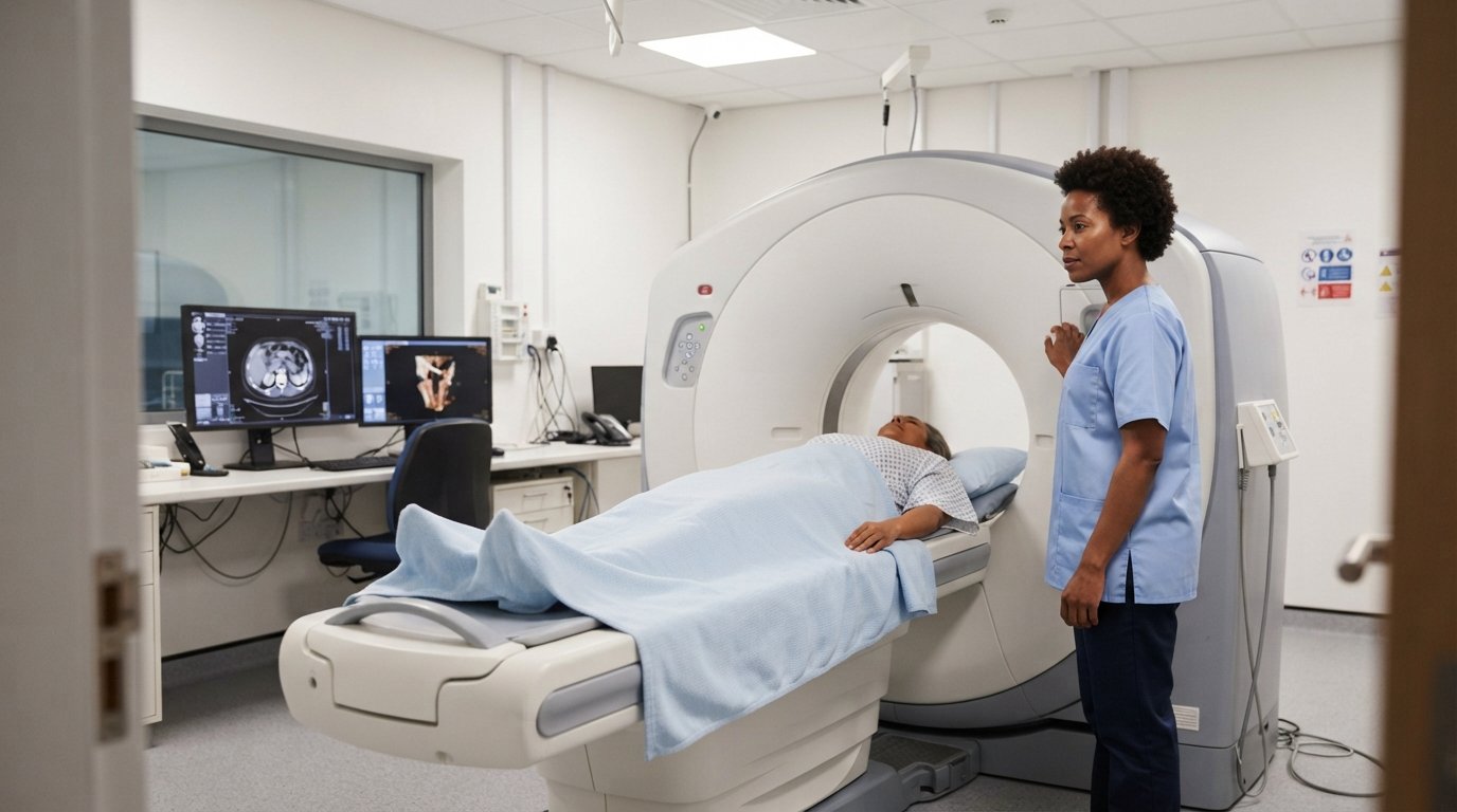
During a CT examination, the patient lies on a motorized table that moves through a large circular opening in the scanner.
An X-ray tube rotates around the body while detectors measure the X-rays that pass through the tissues.
A computer processes these measurements to produce detailed images.
Depending on the examination, CT imaging can show:
- Bones
- Organs
- Blood vessels
- Soft tissues
- Lungs
- Brain
- Abdomen
- Pelvis
- Spine
- Other internal structures
What Is a Scanner Medical Examination?
A medical scanner is another term commonly used for a CT scan.
Doctors may request scanner imaging to investigate symptoms, diagnose injuries, monitor disease, or evaluate the effectiveness of treatment.
CT imaging can be used in emergency departments as well as planned outpatient examinations.
How Does a CT Scanner Work?
A CT scanner combines X-rays and computer technology.
The basic process is:
- The patient lies on the examination table.
- The table moves through the scanner opening.
- An X-ray tube rotates around the body.
- Detectors collect X-ray information.
- A computer processes the collected data.
- The system creates cross-sectional images.
- Radiologists interpret the images.
Modern CT scanners can acquire large amounts of information very quickly.
What Is the Difference Between a Scanner and an X-Ray?
Both CT scanners and conventional X-ray systems use X-rays, but they create images differently.
| Feature | CT Scanner | Conventional X-Ray |
|---|---|---|
| Imaging | Cross-sectional images | Projection image |
| Detail | Generally greater | More limited |
| 3D reconstruction | Possible | Generally not |
| Examination time | Usually short | Usually very short |
| Radiation | Uses ionizing radiation | Uses ionizing radiation |
| Common uses | Detailed evaluation | Bones, chest, dental and other applications |
A CT scan can provide significantly more anatomical information than a single conventional X-ray.
What Is the Difference Between CT and MRI?
CT and MRI are different medical imaging technologies.
CT uses X-rays, while MRI uses magnetic fields and radiofrequency energy.
| Feature | CT Scanner | MRI |
| Main technology | X-rays | Magnetic field + radiofrequency |
| Ionizing radiation | Yes | No |
| Speed | Usually fast | Often longer |
| Bone imaging | Excellent | Useful but different |
| Emergency imaging | Frequently useful | Can be less convenient in some emergencies |
| Soft-tissue contrast | Good | Often excellent |
The appropriate examination depends on the medical question.
What Are the Main Types of Scanner Imaging?
CT imaging can be performed on different areas of the body.
Brain Scanner
A brain CT scan can be used to evaluate:
- Head injuries
- Bleeding
- Stroke-related findings
- Skull abnormalities
- Certain tumors
- Other neurological conditions
CT is particularly valuable in emergency situations because it can be performed rapidly.
Chest Scanner
A chest CT scan can provide detailed images of the lungs, airways, blood vessels, and other structures in the chest.
It may be used to investigate:
- Persistent respiratory symptoms
- Lung nodules
- Infections
- Trauma
- Pulmonary abnormalities
- Certain cancers
Abdominal Scanner
An abdominal CT scan can examine structures such as:
- Liver
- Kidneys
- Pancreas
- Spleen
- Intestines
- Blood vessels
It may help investigate abdominal pain, trauma, inflammation, masses, or other abnormalities.
Pelvic Scanner
A pelvic CT can examine structures in the lower abdomen and pelvis.
Spine Scanner
A spine CT scan can provide detailed information about vertebrae and other bony structures.
It may be used for:
- Trauma
- Fractures
- Degenerative changes
- Bone abnormalities
- Surgical planning
Vascular Scanner
A CT angiography (CTA) examination uses CT imaging, often with contrast material, to visualize blood vessels.
It can be used to examine arteries and other vascular structures.
What Is a Contrast-Enhanced CT Scan?
Some CT examinations use a contrast agent to make certain tissues or blood vessels easier to distinguish.
Depending on the examination, contrast may be administered:
- Intravenously
- Orally
- Through another route appropriate to the examination
Contrast-enhanced CT can improve visualization of specific anatomical structures.
Not every CT scan requires contrast.
What Is CT Contrast?
CT contrast is a substance used to improve the visibility of certain structures during imaging.
Iodine-based contrast agents are commonly used for intravenous CT examinations.
Before contrast administration, medical staff may ask about:
- Previous contrast reactions
- Kidney function
- Medications
- Medical history
- Other relevant risk factors
Always provide complete information to the imaging team.
How to Prepare for a Scanner Examination
Preparation depends on the type of CT scan.
You may be instructed to:
- Avoid eating for a specified period
- Drink or avoid certain fluids
- Remove metal objects
- Wear appropriate clothing
- Take regular medications as instructed
- Arrive early for registration
- Have blood tests when required
If contrast is planned, the imaging center will provide specific preparation instructions.
Always follow the instructions provided by your healthcare provider or imaging facility.
Can You Eat Before a CT Scan?
It depends on the examination.
Some CT scans require fasting, particularly when contrast or specific abdominal imaging protocols are involved.
Other examinations may not require fasting.
Do not assume that every CT scan has the same preparation requirements.
Follow the instructions from your imaging center.
What Happens During Imagerie Scanner?
A typical CT examination is relatively straightforward.
Step 1: Registration and Preparation
You may be asked to complete medical information and confirm the examination.
Step 2: Changing Clothes
Depending on the scan, you may need to change into appropriate clothing.
Step 3: Removing Metal Objects
Metal can interfere with imaging, so you may need to remove jewelry, glasses, or other objects.
Step 4: Positioning
You lie on the CT examination table.
Step 5: Scanning
The table moves through the scanner while the X-ray system collects images.
Step 6: Breath-Holding
For some examinations, you may be asked to hold your breath for a few seconds.
Step 7: Completion
Once the images are obtained, the examination is finished.
Does a CT Scan Hurt?
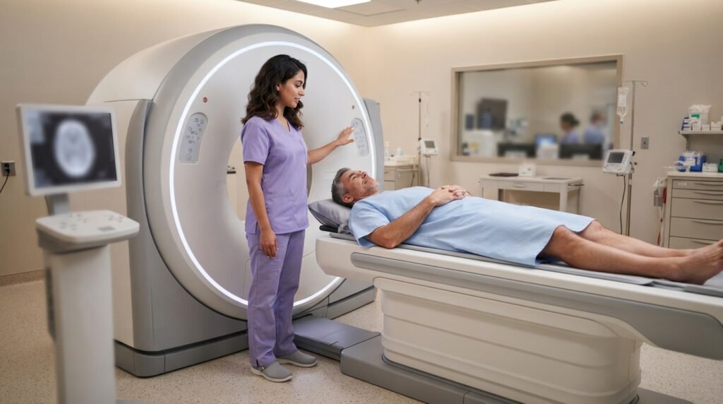
A CT scan itself is generally painless.
You may feel uncomfortable from lying still or maintaining a particular position.
If intravenous contrast is used, you may briefly experience sensations such as:
- Warmth
- A flushing feeling
- Metallic taste
These sensations usually pass quickly.
Tell the medical staff if you experience anything unusual or severe.
How Long Does a Scanner Examination Take?
The actual CT image acquisition can be very fast, sometimes taking only seconds for a particular scan.
However, the total appointment can take longer because of:
- Registration
- Preparation
- Positioning
- Contrast administration
- Additional imaging
- Post-examination procedures
The exact time depends on the type of CT examination.
Is Imagerie Scanner Safe?
CT scans are widely used medical examinations, but they involve ionizing radiation.
The amount of radiation varies depending on:
- Scanner model
- Body area
- Examination protocol
- Patient size
- Number of imaging phases
Medical professionals generally recommend CT when the expected diagnostic benefit justifies the radiation exposure.
Is CT Scan Radiation Dangerous?
Radiation exposure from medical imaging is considered when determining whether an examination is appropriate.
The risk associated with an individual CT examination is generally considered small, but radiation exposure is cumulative over a person’s lifetime.
This is why healthcare professionals aim to use appropriate protocols and avoid unnecessary imaging.
If you are concerned about radiation, discuss the reason for the examination with your healthcare provider.
CT Scan During Pregnancy
Pregnancy is an important consideration before imaging that uses ionizing radiation.
If you are pregnant or think you might be pregnant, tell your doctor and the imaging team before the examination.
They can determine whether CT is appropriate and whether another imaging method may be suitable.
In urgent situations, CT may still be medically necessary.
What Are the Advantages of CT Imaging?
CT has several important advantages.
Fast Imaging
CT can acquire images quickly, making it valuable in emergencies.
Detailed Anatomical Information
It can provide detailed cross-sectional views of the body.
Excellent Bone Visualization
CT is particularly useful for evaluating complex bone structures and fractures.
Blood Vessel Imaging
CT angiography can provide detailed images of blood vessels.
Multiple Reconstruction Views
CT data can often be reconstructed into different planes and 3D representations.
What Are the Limitations of CT Scanning?
Despite its benefits, CT has limitations.
Radiation Exposure
CT uses ionizing radiation.
Contrast Risks
Some examinations use contrast agents that may not be appropriate for everyone.
Limited Soft-Tissue Contrast Compared With MRI
For certain neurological, musculoskeletal, and soft-tissue problems, MRI may provide better contrast.
Incidental Findings
CT scans can reveal unexpected findings that may require additional evaluation.
What Does a Radiologist Do?
A radiologist is a physician trained to interpret medical images.
After a CT examination, the radiologist reviews the images and prepares a report.
The report may describe:
- Normal findings
- Abnormal findings
- Lesions
- Inflammation
- Fractures
- Masses
- Organ changes
- Other relevant observations
The final interpretation should be discussed with the healthcare professional who requested the examination.
How Are CT Scan Results Delivered?
Depending on the medical facility, results may be provided through:
- A written radiology report
- A patient portal
- Digital imaging files
- A healthcare provider
- A follow-up appointment
The time required to receive results varies depending on the facility and urgency of the examination.
Emergency CT scans may be interpreted rapidly, while routine examinations may take longer.
What Does a Normal CT Scan Mean?
A normal CT scan means the radiologist did not identify significant abnormalities within the scope of the examination.
It does not necessarily mean that every possible medical condition has been ruled out.
Some conditions are better evaluated using:
- MRI
- Ultrasound
- Blood tests
- Endoscopy
- Physical examination
- Other diagnostic methods
The meaning of a normal result depends on the symptoms and reason for the scan.
What Does an Abnormal CT Scan Mean?
An abnormal CT result does not automatically mean cancer or another serious disease.
An abnormal finding may represent:
- Inflammation
- Infection
- Injury
- Benign growth
- Normal anatomical variation
- Scar tissue
- Fluid
- Degenerative changes
- A condition requiring further investigation
Your healthcare provider can explain what a specific finding means in your individual situation.
What Is 3D Scanner Imaging?
Modern CT scanners can collect data that allows computers to create three-dimensional reconstructions.
3D CT images can be particularly useful for:
- Complex fractures
- Surgical planning
- Vascular anatomy
- Certain tumors
- Anatomical visualization
The primary diagnostic information often comes from the underlying cross-sectional images, with 3D reconstructions providing additional visualization.
What Is a CT Scan With Contrast?
A CT scan with contrast uses a contrast agent to improve visualization of selected tissues or blood vessels.
Contrast-enhanced imaging can be useful when doctors need to distinguish structures that may otherwise appear similar.
The decision to use contrast depends on the clinical question.
What Should You Tell the CT Staff?
Before your examination, tell the imaging team about relevant information such as:
- Pregnancy or possible pregnancy
- Previous contrast reactions
- Kidney problems
- Current medications
- Allergies
- Implanted devices
- Relevant medical conditions
The exact information required depends on the examination.
Imagerie Scanner vs Ultrasound
CT and ultrasound use different technologies.
Ultrasound uses high-frequency sound waves and does not use ionizing radiation.
CT uses X-rays and computer processing.
Ultrasound can be especially useful for:
- Pregnancy imaging
- Gallbladder evaluation
- Certain abdominal examinations
- Soft tissues
- Blood-flow assessment using Doppler
CT can provide broader cross-sectional imaging and is often more useful for complex anatomical evaluation.
Imagerie Scanner vs X-Ray
A conventional X-ray is usually quicker and uses a lower radiation dose than CT, but it provides less detailed cross-sectional information.
CT is generally selected when more detailed anatomical information is needed.
The choice depends on the clinical question.
Frequently Asked Questions
1. What does imagerie scanner mean?
Imagerie scanner generally refers to medical imaging performed using a CT scanner, also called computed tomography.
2. What is a scanner in medical imaging?
A medical scanner or CT scanner uses X-rays and computer processing to produce cross-sectional images of the body.
3. Is a CT scan the same as an MRI?
No. CT uses X-rays, while MRI uses magnetic fields and radiofrequency energy.
4. Is a CT scan painful?
The scanning process itself is generally painless. Some people may experience temporary sensations when intravenous contrast is administered.
5. How long does a CT scan take?
Image acquisition can be very fast, but the complete appointment may take longer because of preparation, positioning, contrast administration, and other procedures.
6. Do all CT scans require contrast?
No. Many CT examinations can be performed without contrast. The decision depends on the medical purpose of the scan.
7. Can you eat before a scanner examination?
Preparation varies. Some examinations require fasting, while others do not. Follow the specific instructions provided by the imaging facility.
8. Does a CT scan use radiation?
Yes. CT uses ionizing X-rays to create images.
9. Is CT scan radiation dangerous?
CT involves radiation exposure, so it should be performed when medically appropriate. Healthcare professionals consider the potential benefits and risks when recommending the examination.
10. Can CT detect cancer?
CT can identify masses, tumors, enlarged lymph nodes, and other abnormalities that may be associated with cancer. Additional tests may be necessary to determine the exact diagnosis.
11. Can a CT scan detect inflammation?
Yes. CT can show certain signs of inflammation, infection, swelling, fluid collections, and other abnormalities.
12. Is CT better than MRI?
Neither is universally better. CT is fast and excellent for many bone, lung, emergency, and vascular applications, while MRI often provides superior soft-tissue contrast for certain conditions.
13. Can you drive after a CT scan?
For many CT examinations without sedation, people can generally return to normal activities. If sedation or another procedure is involved, different instructions may apply.
14. When will I receive my CT scan results?
The timing varies by facility and examination urgency. Emergency studies may be reviewed quickly, while routine studies can take longer.
Final Thoughts
Imagerie scanner is an important part of modern medical imaging and generally refers to CT or computed tomography imaging. CT scanners use X-rays and computer processing to create detailed cross-sectional images of the body. CT can be used to examine the brain, chest, abdomen, pelvis, spine, bones, blood vessels, and many other structures. It is particularly valuable when rapid and detailed imaging is needed.
Although CT uses ionizing radiation, medical professionals carefully consider the expected diagnostic benefit when recommending the examination. Contrast may also be used when additional visualization of certain tissues or blood vessels is required.
If your doctor has recommended a scanner examination, follow the preparation instructions provided by the imaging center and inform the medical team about pregnancy, allergies, previous contrast reactions, medications, and relevant medical conditions. Most importantly, remember that a CT report should be interpreted in the context of your symptoms, medical history, physical examination, and other diagnostic information by your healthcare professional.
Related Post
- Indianapolis Police Scanner – Complete Guide to Live Police Radio, Frequencies, Apps & Online Listening!
- Kenosha Scanner – Complete Guide to Kenosha Police Scanner, Fire Scanner, Frequencies, Apps, and Online Listening!
- Lapd Scanner – Complete Guide to Los Angeles Police Scanner Frequencies, Apps, and Public Safety Radio!
About the Author